

The "Keyhole Problem": Why Your Pupil Limits Your Doctor's View
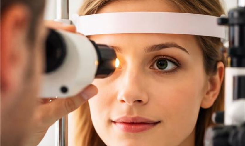
Imagine trying to inspect an entire room just by peeking through a keyhole. You’d only see a tiny fraction of what’s inside, no matter how hard you looked. This is exactly the challenge your eye doctor faces during a standard, undilated eye exam. Your pupil—the small black circle in your eye's center—acts like that keyhole, offering only a very limited view of the vast area behind it.
How Dilation "Opens the Door" to Your Eye's Inner Health
To solve the keyhole problem, your doctor uses special mydriatic eye drops. These drops work by temporarily relaxing the muscle in your iris—the colored part of your eye—that’s responsible for shrinking your pupil. By preventing the pupil from constricting in the bright light of the exam, the drops essentially prop the "door" to your eye wide open, making a thorough inspection possible.
What Your Eye Doctor Can Actually See with Dilation
With the door to your eye open, your doctor gets a clear, panoramic view of the retina. Think of the retina as the light-sensitive sensor at the very back of your eye, like the film in a camera. It’s the tissue that captures everything you see and begins turning it into information your brain can understand.
During a comprehensive dilated eye exam, your doctor will closely inspect your optic nerve. This is the vital “data cable” that connects the retina to your brain. Checking this structure is the most effective way to spot early signs of glaucoma, a disease that can quietly damage the nerve and lead to permanent loss of your peripheral, or side, vision.
Your doctor also examines the macula, a tiny, specialized part of the retina responsible for your sharp, high-definition central vision. It’s what allows you to read, drive, and recognize faces. This is where they look for signs of macular degeneration. Furthermore, the full retinal view allows them to spot changes in tiny blood vessels, which is critical for the early detection of diabetic retinopathy.
Is Dilation Always Necessary? Optomap vs. Dilation
With advancing technology, you might wonder if getting your eyes dilated is necessary. Many clinics now offer digital retinal imaging, like an Optomap, which takes a panoramic 2D photograph of your retina. This provides an excellent wide-angle snapshot of your eye’s health and is a fantastic screening tool.
Your Survival Guide: Preparing For a Dilated Eye Exam
Knowing what to expect can turn a dilated exam from a hassle into a smooth process. The most common concerns are the duration of the effects and whether you can drive afterward. Because driving is unsafe while your pupils are dilated, you must arrange for another way home.
- Bring dark sunglasses (even on a cloudy day!).
- Arrange for a driver to take you home.
- Plan to rest your eyes; avoid computer work or reading for a few hours.
- Allow 4-6 hours for the main effects to wear off.
The Small Price for a Lifetime of Sight
That blurry inconvenience now makes sense. The "keyhole" of your pupil limits a doctor's view, and dilation is the only way to open the door for a complete health check. Instead of a mysterious requirement, the procedure is a logical, preventative step.